Staging of Breast Cancer helps us to know the severity of the breast cancer based on how far it has spread. Staging is done based on the size of tumour, presence of enlarged lymph nodes and the spread beyond lymph nodes to other parts of the body. Based on this there are 4 stages of breast cancer. Stages I & II are generally called as Early Breast Cancer in which the tumour is generally less than 5 cms in size and the spread has been limited to the lymph nodes in the same side axilla
Non palpable breast cancers are generally diagnosed by screening. This is done by mammograms taken once every year above the age of 40 years. Women who have a strong family history may take mammograms above the age of 30 years. As the breast is dense in the younger age group, mammograms maybe coupled with ultrasound to find out the presence of non palpable breast cancers. Finding the breast cancer in this stage itself will be very good as the survival rates for these type of cancers is very high.
Palpable breast cancers are diagnosed with core needle biopsy which is done in the clinic itself. This can be done with the help of ultrasound. A 5 mm small incision is made over the breast. A core needle is inserted into the breast and a small amount of tissue is taken along with the needle. This tissue taken out is then sent for biopsy. The biopsied tissue is analysed by a pathologist who sees the tissue under a microscope and sees whether the tissue is a cancer or not. If there is cancer, further immunohistochemical markers such as hormone receptors like estrogen receptor and progesterone receptors are looked for in the tissue biopsied. This gives us a lot of information for further treatment.
The investigations done can be broadly divided into*
Mammogram, Mammogram with Ultrasound, Ultrasound Guided Core Needle biopsy
Fine Needle Aspiration Cytology (FNAC) & Sentinel Lymph node Biopsy (SLNB) for lymph node spread to the axilla or armpit, Chest X Ray or CT chest for spread to the lungs, Ultrasound Abdomen or CT Abdomen for spread to the abdomen
Blood Investigations, ECG, Chest X ray, Echocardiogram
Early breast cancer is treated initially by surgery. If Breast Conservative therapy or Therapeutic Mammaplasty had been done the patient would require radiotherapy after chemotherapy. After surgery, the removed breast cancer is sent to the pathologist who examines the tissues under the microscope and confirms the diagnosis, tells us about the histology, grade of the tumour and whether the removed breast tissue has estrogenreceptors, progesterone receptors and Her2 neu receptors. The information provided by the pathologist is necessary to know whether we need to give chemotherapy, radiotherapy and hormone therapy.
The surgical modalities for breast cancer can be divided into those procedures which remove the cancer tissue from the breast and those surgical modalities which remove the lymph nodes in the axilla. We would need to choose one procedure from each modality. The procedure that is chosen for each patient will depend on the size of the tumour, stage of the tumour, size of the breast, presence of multiple lesions and patient preference. The various surgical procedures to remove the cancer tissue from the breast are
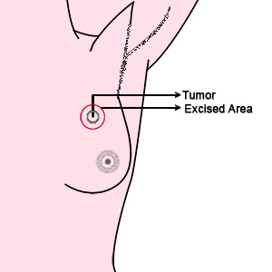
This can be done only for a small tumour in a large breast where in we remove the breast cancer along with a small margin of tissue. If there are multiple lesions in the breast this procedure can not be done. The patient would definitely need radiotherapy after this procedure. By doing this we can conserve the breasts.(To know more about Breast Conservative Therapy please click here)
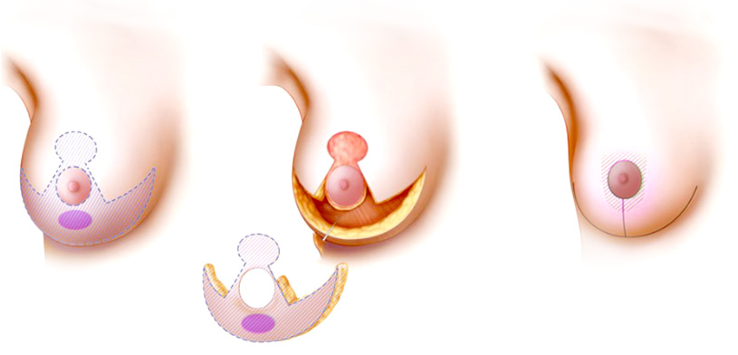
This procedure is done for slightly larger tumours in a large breast where in we remove a part of the breast and use breast reduction plastic surgical techniques to reduce the size of the breast. This breast would definitely need radiotherapy after the procedure.(To know more about Therapeutic Mammaplasty please click here)
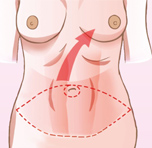
Here the whole breast is removed. This is done for small breasts, multiple tumours in the breast and breast cancers which present in an advanced stage. We offer the full gamut of reconstruction possibilities for the patient. The best method of reconstruction would be autologous reconstruction where we take the patients own tissues such as the tummy, inner thighs, back or buttocks to make a new breast. Reconstruction of the breast is best done at the same time of the mastectomy or removal of the breast as the patient wakes up with a new breast, surgery is done in one operation and the cosmetic result of a breast done at the same time of removal of the breast is better than when reconstructed at a later date.
Here the whole breast is removed. Some women may not want any form of
reconstruction of the breast and would like to remain flat chested. They can use
prosthesis in their bras if necessary or they can get their breasts
reconstructed after some time.
Surgical procedures to address the lymph nodes in the axilla
The first place that breast cancer tends to spread is the lymph nodes in the axilla or the armpit. The procedures done to address the spread to the axilla are
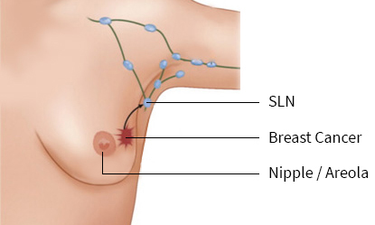
Sentinel lymph node is the first lymph node that drains the breast cancer. It is assumed that if this lymph node is biopsied and no tumour is found then more radical procedures such a axillary lymph node dissection can be avoided. However if tumour is found in the lymph node biopsied, a complete axillary lymph node dissection needs to be done (To know more about sentinel lymph node biopsy please click here)
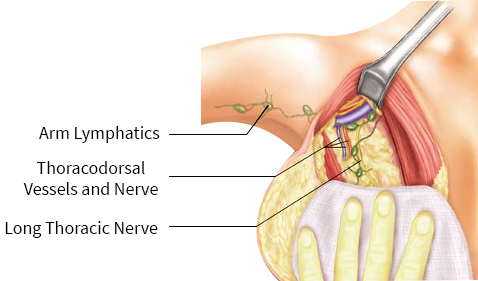
Axillary lymph node dissection is done when there is a lymph node which is palpable in the axilla or if sentinel lymph node biopsy is positive. Here the entire group of lymphatics in the axilla is removed. (To know more about axillary dissection please click here)
Chemotherapy drugs are injected intravenously. As the drugs go to all parts of the body it helps to kill the cancer cells which have spread to any other part of the body. Chemotherapy is necessary when the tumour is above 2 cms in size, has spread to a lymph node, absent estrogen receptors, Her 2 neu positive, unfavourable histology and high grade tumours. This is usually given within 6 weeks after surgery.
Radiotherapy is done when surgical modalities such as Breast Conservative Therapy and Therapeutic Mammaplasty are chosen as the preferred surgical modality. It is also necessary when the tumour is above 5 cms in size, has spread to a lymph node and has positive or close margins. This is generally done after surgery and chemotherapy have been completed.
After core needle biopsy and surgery, the tissues are sent for biopsy and the pathologist lets us know if there are receptors for hormones such as estrogen and Her 2 neu receptors. If the hormone receptor are present then hormone therapy is given. This consists of oral medicines taken for around 5 to 10 years. This is done after surgery, chemotherapy and radiotherapy .
We at Ganga Hospital will be happy to answer any doubts that you may have regarding treatment for breast cancer, breast reconstruction or for cosmetic surgery for the breast. Please enter your mobile number and your email ID so that we can reply back to you. You can scan and attach your reports or photos which would enable us to make a decision. Alternatively you can whats app your doubts to the Ganga Breast Care Unit number +91 9952617171. The corresponding doctor would respond to you within 24 hours

